
Models and Descriptors for typical and pathological development of Gyrification – J. Lefèvre
 This project is dedicated to new theoretical approaches to cerebral development that we apply to concrete neuroscientific and clinical issues in medical imaging. A multi-disciplinary approach is proposed to study early sulcal morphogenesis. It is based on valuable data sets of MRI images from fetuses, preterm newborns, infants and children. Two main methodological research line are investigated. In the first one we are looking for quantitative descriptions of cortical anatomies in the equivalent of the “image” and “Fourier” domain, adapted to specific geometrical representations. In the second one we develop computational models of folding process based on a coupled mechanical and reaction-diffusion models.
Shape analysis and the quantification of cortical gyrification – J. Lefèvre, O. Coulon, H. Rabiei
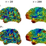 In this project we investigate the theoretical aspects of spectral shape analysis in order to characterize cortical gyrification. In particular we develep a window Fourier transform technique for meshed surfaces. Measurements of sulcal gyrification are derived from this WFT. This work is done in close collaboration with F. Richard (Institut de Mathématiques de Marseille) and is funded by the Labex Archimède.
Morphometry of cortical sulci – O. Coulon, J. Lefèvre
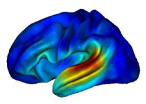
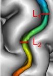 We developped several methods that aim to quantify the morphology of sulci. These methods have been applied in particular to the Central Sulcus of humans (collaborators: M.Cykowski, University of Texas in San Antonio; M.Dojat, Grenoble Institute of Neuroscience; J.-F. Mangin, Neurospin Center; H. Siebner, University Medical Center Freiburg; S. Klöppel, Danish Research Center for Magnetic Resonance) and a variety of non human primates (collaborator: W. Hopkins, Yerkes National primate Research Center) . Asymmetries of depth have been characterized and related to handedness, and we proposed a reliable method to automatically detect the position of the ‘hand knob’ and correlate this position with handedness. In a recent collaboration with F. Leroy (Neurospin Center) these methods were applied to the Superior Temporal Sulcus and revealed a very stable inter-hemispheric asymmetry of this sulcus.
Sulcal pits as markers of gyrification and development – G. Auzias, O. Coulon, C. Deruelle (SCALP team), L. Brun, J. Lefèvre, S. Takerkart, I. Kaaltenmark, A. Pron
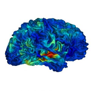
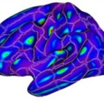 Sulcal pits, the points at which sulcal depth is reaching a local maximum, are thoughts to be very good markers of cortical gyrification, and their link with cortical development makes them good candidate features to characterize developmental trajectories. In this line of research we investigate the algorithmic and conceptual aspects of sulcal pits characterization, and we propose methods to use sulcal pits for cortical morphometrics and building models of normal development. C. Deruelle also investigate their use to find biomarkers of developmental pathologies such as autism.
Neuroanatomical correlates of voice perception and correlation between anatomy and function – O. Coulon, P. Belin (BaNCo team) C. Bodin, S. Takerkart
 In this project we study functions associated with voice perception, a central topic of research for P. Belin, their localization around the Superior Temporal Sulcus, and their correlation with the STS morphology. A large database acquired with human subjects is being analyzed and further research will aim at using non human primates in order to provide inter-species comparison and understand further the specificities of voice perception and communication.
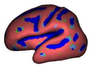
MarsAtlas: a cortical parcellation atlas for the analysis of MEG functional data – A. Brovelli (CoMCo team), O. Coulon, G. Auzias
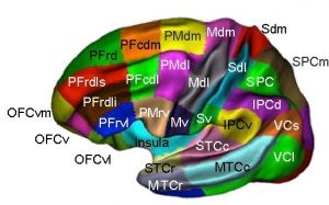 Based on the same macroanatomical model than the HipHop parameterization model, we developped a cortical surface parcellation technique that can be used to analyze functional data. We investigate the level of functional segregation that can be defined with such model and apply it to MEG data analysis in collaboration with A. Brovelli (CoMCo team, INT).
Functional data analysis in a clinical context – O. Coulon, S. Takerkart, L. Thiébaut
 In collaboration with Olea Médical, we investigate the possibility to use resting-state functional connectivity in a clinical context. We aim to produce efficient data analysis methods that comply to the constraints of the clinical environment.
White matter morphometry and rehabilitation – L. Velly
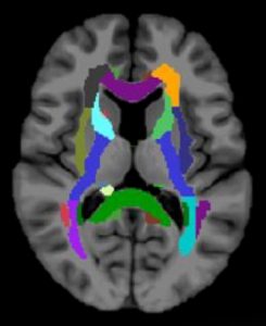 In this project we are investigating white matter measurements from diffusion imaging as biomarkers of rehabilitation in emergency patghologies such as head trauma, cardiac arrest, or stroke.